Breakthrough Technique Reveals Unseen Features of Synaptic Organization in the Brain
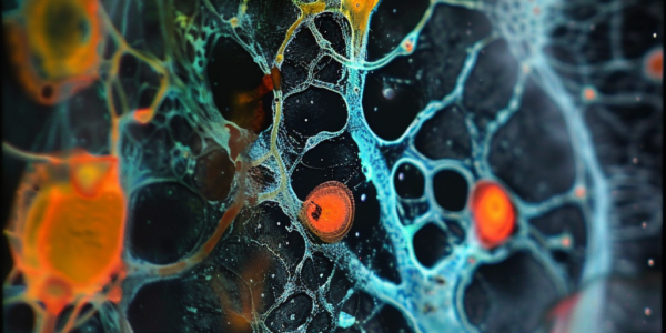
Researchers have developed a cutting-edge technique for high-throughput volumetric mapping of synaptic transmission, revealing new insights into synaptic organization in the brain. The study, ‘High-throughput volumetric mapping of synaptic transmission,’ published in Nature Methods, employed a novel approach using Bessel-droplet foci for high-resolution imaging of synapses. This groundbreaking research offers valuable implications for neuroscience and beyond.
Revolutionizing Neuroscience: MIT Researchers Develop Technology for 3D Imaging of Entire Human Brain Hemispheres
Exciting advancements in neuroscience research have been made as a team of researchers from MIT successfully developed a technology pipeline for 3D imaging of entire human brain hemispheres at subcellular resolution. This groundbreaking achievement opens up new possibilities for studying the intricacies of the human brain in health and disease, offering new insights into neurodegenerative diseases like Alzheimer’s.
Study Shows Lung Cancer Screening Saves Lives
Recent study shows that lung cancer screening can save lives, with significantly higher rates of early disease diagnosis and lower death rates among screened patients. National efforts to promote screening initiatives are highlighted, emphasizing the importance of early detection in improving outcomes for lung cancer patients.
Transparent Skull Implant Enables Revolutionary Brain Imaging Technology
Researchers at the Keck School of Medicine of USC and Caltech have developed a new brain imaging technique using a transparent ‘window’ in a patient’s skull. This innovative approach, demonstrated in a proof-of-concept study, utilizes functional ultrasound imaging to record brain activity. Led by Dr. Charles Liu, the study shows promising implications for patient monitoring and a deeper understanding of brain function, particularly in individuals with neurological disabilities. This groundbreaking research offers new possibilities for diagnosis and treatment in patients with serious head injuries.
Optical Coherence Tomography: A Game-Changer in Detecting Ureteral Injuries During Pelvic Surgery
Discover how optical coherence tomography (OCT) endoscopy is revolutionizing the detection of ureteral injuries during pelvic surgery. Unlike traditional methods, OCT provides real-time visualization of tissue damage, enabling prompt interventions and improving patient outcomes. Learn how this advanced imaging technology is enhancing surgical procedures and shaping the future of medical care.
Chest CT Scans as a Tool for Predicting Low Bone Mineral Density
A recent study published in Clinical Interventions in Aging highlights the potential of utilizing chest CT scans as an alternative to DEXA for assessing bone health. By analyzing CT scans for vertebral compression fractures, healthcare providers can detect and manage low bone mineral density more effectively. The study underscores the benefits of incorporating opportunistic chest CT scans in assessing bone health, ultimately improving patient outcomes.
Study Finds Potential Negative Implications of Large Language Models in Breast Imaging Classification
A recent study published in Radiology highlights the potential negative implications of using large language models like GPT-4 and Google Gemini in breast imaging classification. While these AI models have shown promise in certain tasks, they may fall short in more complex medical reasoning. The study compared the performance of LLMs with board-certified breast radiologists in assigning BI-RADS categories, revealing a lack of strong agreement. Lead author Dr. Andrea Cozzi stresses the importance of evaluating the limitations of generic LLMs, especially in scenarios where medical reasoning is critical. The findings emphasize the need for better regulation of LLMs in medical settings to ensure accurate classification of imaging reports and improve patient care.
New Imaging Method Enhances Precision in Prostate Cancer Treatment
Groundbreaking imaging method utilizing lead-212 (212Pb) offers new hope for prostate cancer patients. The SPECT/CT acquisition technique enhances precision in treatment by accurately detecting radiopharmaceutical biodistribution. This innovative approach revolutionizes cancer treatment by providing valuable insights into drug biodistribution and pharmacokinetics, promising more personalized therapies. Published in The Journal of Nuclear Medicine, the first-in-human images showcase the efficacy of the technique in improving patient outcomes through tailored therapies.
AI Revolutionizes Retinal Imaging
Artificial intelligence has revolutionized retinal imaging, making it 100 times faster and improving image contrast 3.5-fold. This advancement is a game-changer for evaluating age-related macular degeneration and other retinal diseases, offering researchers a more effective tool for early detection and treatment.
A.I. Revolutionizing Breast Cancer Screening
Artificial intelligence (A.I.) is revolutionizing breast cancer screening, offering the potential to enhance accuracy and detect cancer earlier. A.I. models can detect subtle patterns in mammograms that may be challenging for human radiologists to differentiate, sparking excitement within the medical community. However, concerns remain about the effectiveness of A.I. tools across diverse patient populations and their impact on breast cancer survival rates.


