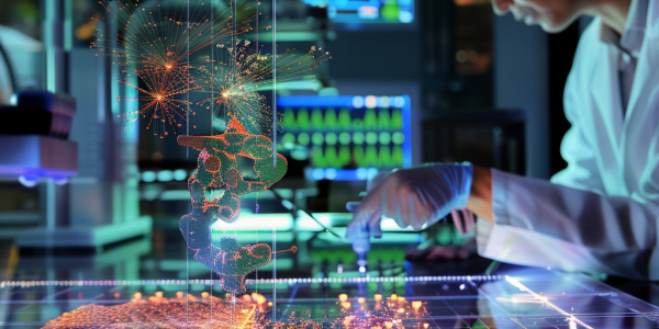
MIT Researchers Unveil Breakthrough in Metabolic Imaging Techniques
A groundbreaking study from MIT introduces a novel metabolic imaging technique that significantly enhances penetration depth, allowing for clearer and more detailed visualization of living tissues. This innovative approach accelerates imaging processes and eliminates the need for tissue preprocessing, paving the way for advancements in cancer research, drug discovery, and more.
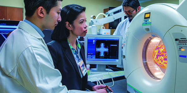
Breakthrough MRI Technology Predicts Ovarian Cancer Treatment Response
A groundbreaking study from the University of Cambridge reveals a revolutionary MRI-based imaging technology, hyperpolarized carbon-13 imaging, that can predict ovarian cancer tumor responses to treatment. This innovative method enhances MRI signals by up to 10,000 times, enabling rapid assessments of treatment effectiveness, distinguishing between tumor subtypes, and personalizing cancer care. With the ability to evaluate responses to chemotherapy within 48 hours, this advancement promises to transform ovarian cancer treatment and improve patient outcomes.
Mica AI Partners with Baptist Health to Revolutionize Breast Cancer Diagnosis
Mica AI Medical Ltd. is revolutionizing breast cancer diagnosis with its advanced software that enhances mammogram analysis. Recently partnering with Baptist Health South Florida, Mica AI aims to improve accuracy in detecting breast cancer, reducing false positives and negatives. This collaboration marks a significant entry into the U.S. healthcare market, showcasing the growing role of artificial intelligence in medical diagnostics and the potential for better patient outcomes.
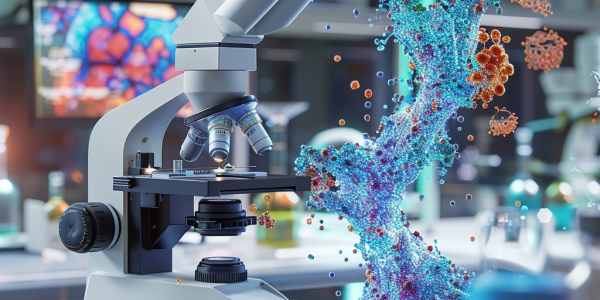
MIT Researchers Unveil Groundbreaking 20-Fold Tissue Expansion Technique for High-Resolution Imaging
MIT researchers have developed a groundbreaking 20-fold tissue expansion technique that allows high-resolution imaging of nanoscale structures using standard light microscopes. This innovative method democratizes access to advanced biological imaging, reducing costs and enabling more laboratories to conduct detailed studies of cellular components, such as synapses and proteins. Published in Nature Methods, this technique promises to transform biological research and open new avenues for scientific discovery.
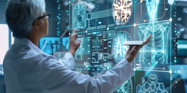
Revolutionizing Radiology: AI Enhances Diagnostics and Patient Care
The integration of artificial intelligence (AI) in healthcare, particularly in radiology, is transforming diagnostics. Companies like Qure.ai are leading this change with advanced AI tools that enhance efficiency and accuracy, significantly improving patient outcomes. AI’s ability to swiftly analyze medical images, such as X-rays and CT scans, is crucial in areas with limited access to radiologists, offering timely diagnoses and reliable results. As AI continues to evolve, its collaboration with healthcare professionals promises to redefine standards of care in medical imaging.

Revolutionary Noninvasive Imaging Technique Enhances Monitoring of Cardiovascular Health Post-Meal
A groundbreaking study from Boston University and Harvard Medical School introduces a noninvasive imaging technique, Spatial Frequency Domain Imaging (SFDI), to monitor cardiovascular health post-meal. This innovative method tracks how different meal compositions, particularly low-fat and high-fat diets, affect tissue properties and hemodynamics, providing valuable insights for proactive heart health management.
Revolutionary NIR Thermometer Enhances Temperature Measurement Accuracy
Recent advancements in thermal imaging technology from the University of Houston promise to revolutionize temperature measurement in military and medical applications. A new non-contact thermometer utilizing near-infrared spectroscopy offers precise readings without reliance on emissivity values, addressing limitations of traditional methods. This innovation enhances diagnostic capabilities and operational efficiency, paving the way for breakthroughs in various fields.
Revolutionizing Prostate Cancer Diagnosis with PSMA-PET Imaging
Discover the future of prostate cancer diagnosis with PSMA-PET imaging, as Dr. Brian T. Helfand highlights advancements in imaging technologies that promise earlier detection and personalized treatment strategies. Learn how innovative radiotracers and a deeper understanding of tumor biology are set to revolutionize urologic oncology.
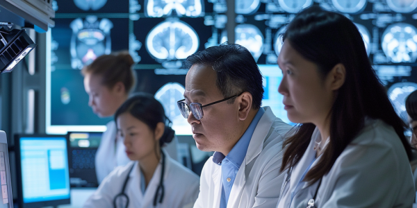
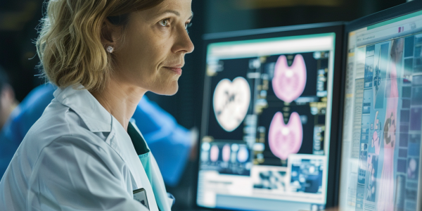
Sydney Neuroimaging Centre Secures Funding for AI-Powered Brain Disease Analysis Software
Sydney Neuroimaging Analysis Centre has secured significant government funding and regulatory approval for its innovative AI-powered software to analyze and monitor brain diseases. The A$3.75 million funding from ANDHealth+ will advance the commercialization of its AI-enabled medical imaging software, iQ-solutions, offering precise analysis of brain structures from MRI scans. SNAC is also developing tools like Torana and VeriScout for medical scan management and automatic detection of cerebral hemorrhage. With global market clearances, SNAC is set to make a significant impact in the $250 million brain image analysis market.

AI Revolutionizing Women’s Healthcare with Generative AI (GenAI)
Discover how Artificial Intelligence (AI), especially generative AI (GenAI), is revolutionizing women’s healthcare by bridging historical gaps in research and development processes. Learn how AI technologies are addressing disparities in healthcare solutions for women by leveraging physiological and hormonal differences to improve treatment outcomes.