Scientists Create Organized Stem Cell Culture Model Resembling Human Embryonic Brain and Spinal Cord
In a groundbreaking development, a team of engineers and biologists at the University of Michigan, the Weizmann Institute of Science, and the University of Pennsylvania has successfully created an organized stem cell culture model that resembles all three sections of the embryonic brain and spinal cord. This achievement marks the first time a model has been able to produce a full representation of the early stages of the human central nervous system.
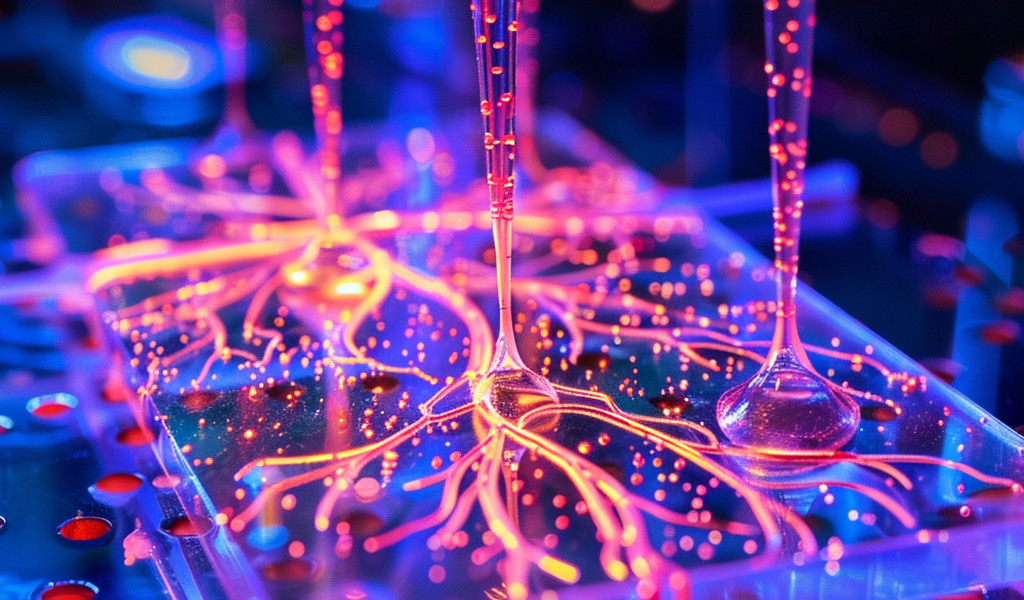
The model, a neural tube organoid derived from human pluripotent stem cells (hPSCs), is grown inside a microfluidic device and requires daily replenishment of fresh media. Jeyoon Bok, a PhD candidate at the Department of Mechanical Engineering, played a crucial role in changing the fluid that feeds the stem cells and guiding their differentiation into the model of the early human central nervous system.
According to Jianping Fu, PhD, a professor of mechanical engineering at the University of Michigan, models like this will pave the way for fundamental research to understand the early development of the human central nervous system and its implications in various disorders.
The research, published in Nature under the title ‘A Patterned Human Neural Tube Model Using Microfluidic Gradients,’ has the potential to revolutionize the study of neurological and neuropsychiatric diseases. Guo-Li Ming, PhD, and Hongjun Song, PhD, both Perelman professors of Neuroscience at UPenn, were instrumental in developing protocols for growing and guiding the cells, as well as characterizing the structural and cellular characteristics of the model.
While current human brain and spinal cord organoids are utilized for studying neurological and neuropsychiatric diseases, they often fail to mimic the complete central nervous system and lack organization. Specifically, they struggle to recapitulate neural patterning along both rostral-caudal (R–C) and dorsal-ventral (D–V) axes in a three-dimensional (3D) tubular geometry, a key feature of NT development.
In contrast, the newly developed model successfully replicates the development of all three sections of the embryonic brain and spinal cord simultaneously, a feat that has not been achieved before.