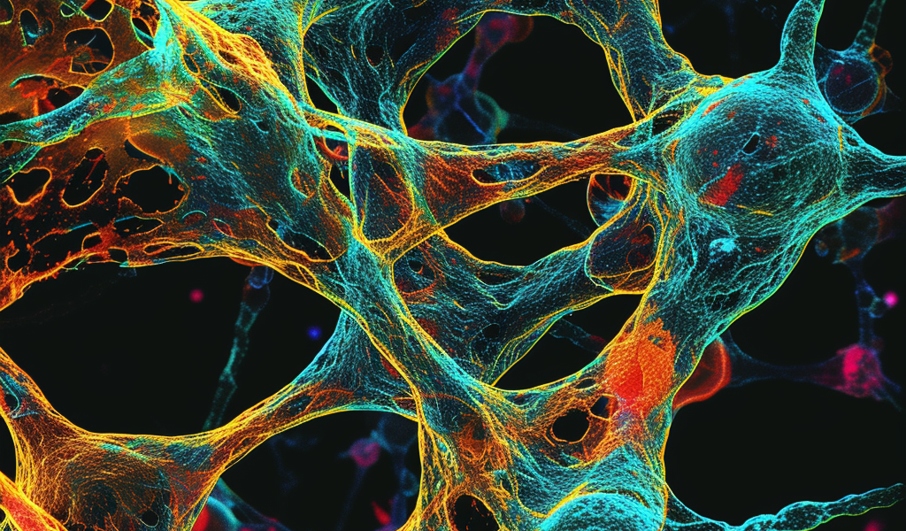
Researchers Capture High-Resolution Images of Early Microtubule Formation in Human Cells
Researchers at the Center for Genomic Regulation (CRG) in Barcelona and the Spanish National Cancer Research Center (CNIO) in Madrid have achieved a groundbreaking feat by capturing the world’s first high-resolution images of the earliest moments of microtubule formation inside human cells. The findings, published in the journal Science, hold immense potential for advancing treatments for various diseases, including cancer and neurodevelopmental disorders.
ICREA Research Professor Thomas Surrey, the main co-author of the study, expressed the significance of the research, stating, ‘Microtubules are critical components of cells, but all the images we see in textbooks describing the first moments of their creation are models or cartoons based on structures in yeast. Here we capture the process in action inside human cells. Now that we know what it looks like, we can explore how it’s regulated. Given the fundamental role of microtubules in cell biology, this could eventually lead to new therapeutic approaches for a wide range of disorders.’
Microtubules, which are tubes made of proteins, play a crucial role in the functioning of cells. They act as bridges or roads, facilitating the movement of substances within the cell and contributing to the cell’s shape. Additionally, microtubules are essential for cell division and serve as transport highways in neurons.
The construction of microtubules involves a complex process carried out by a large assembly of proteins known as the gamma-tubulin ring complex (γ-TuRC). This assembly lays down tiny building blocks called tubulins in a specific order, a process known as microtubule nucleation. Once the foundation is set, tubulins are added to extend the microtubule structure.
It is crucial for microtubules to be made of thirteen different rows of tubulins for the cell to function correctly. However, researchers were puzzled when they discovered that human γ-TuRC exposes fourteen rows of tubulins, contrary to their expectations. The high-resolution images captured in this study provide valuable insights into this discrepancy and pave the way for further exploration of microtubule formation.


