Groundbreaking Discovery in Neuroscience Reveals Unseen Details of Human Brain Structure
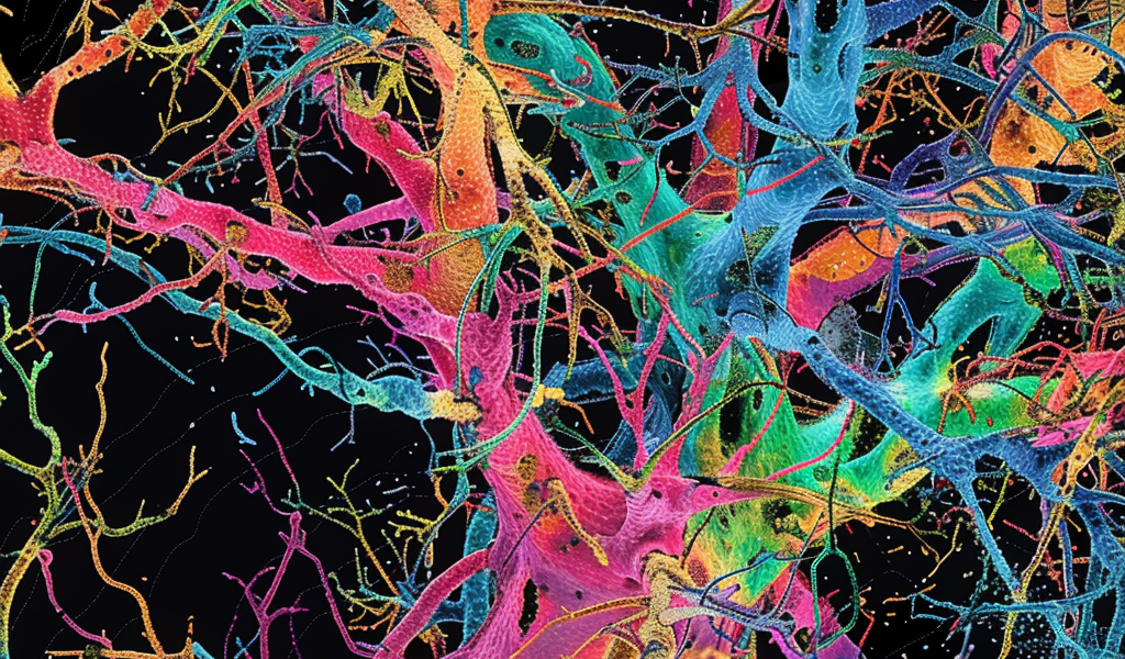
Researchers have recently made a groundbreaking discovery in the field of neuroscience, unveiling unseen details of the human brain’s structure. A team of researchers, led by Dr. Jeff Lichtman at Harvard University and Dr. Viren Jain at Google Research, utilized electron microscopy to image a cubic millimeter-sized piece of human brain tissue at an incredibly high resolution.
The tissue, extracted from the cerebral cortex of a patient undergoing epilepsy surgery, was meticulously sliced into over 5,000 sections, with each section then imaged using electron microscopy. This extensive process resulted in a massive amount of data, approximately 1.4 petabytes, allowing the researchers to create a detailed 3D reconstruction of nearly every cell in the sample.
Analysis of the reconstructed cells unveiled a total of more than 57,000 cells, predominantly neurons and glial cells. Glial cells, which support neurons, outnumbered neurons by a ratio of 2-to-1. Oligodendrocytes, a type of glial cell providing structural support and electrical insulation to neurons, were among the most common in the sample. Additionally, the sample contained approximately 230 mm of blood vessels.
The researchers discovered novel structural details through the reconstruction process, particularly in triangular cells located in the deepest layer of the cerebral cortex. Many of these cells exhibited two distinct orientations, with mirror images of each other, the significance of which remains a mystery.
Utilizing machine learning, the team identified synapses, the crucial junctions facilitating communication between neurons. This groundbreaking research, funded by the NIH and published in Science, sheds new light on the intricate connections within the human brain and provides a valuable resource for further studies.
This study marks a significant step forward in unraveling the complexities of the human brain and underscores the importance of high-resolution imaging techniques in advancing our understanding of brain structure and function.


