Groundbreaking Bioluminescence Imaging Technique Revolutionizes Study of Oxygen Movement in the Brain
A groundbreaking study has introduced a cutting-edge bioluminescence imaging technique that allows scientists to observe the movement of oxygen in the brain in real-time. This innovative method, inspired by firefly proteins, offers detailed insights into oxygen distribution and has the potential to revolutionize our understanding of brain hypoxia and related diseases.
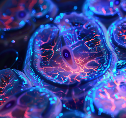
The new imaging technique, unveiled in a recent issue of Science, has provided researchers with visually striking and highly detailed images of oxygen movement in the brains of mice. By enabling continuous monitoring of changes in oxygen concentration across a wide area of the brain, this method has shed light on previously undetected areas of temporary hypoxia, which are indicative of changes in blood flow and can lead to neurological deficits.
According to Maiken Nedergaard, co-director of the Center for Translational Neuromedicine at the University of Rochester and the University of Copenhagen, this breakthrough research has the potential to enhance our understanding of conditions such as hypoxia resulting from strokes or heart attacks. Furthermore, it has revealed that sedentary lifestyles may be linked to an increased risk of Alzheimer’s disease due to the presence of ‘hypoxic pockets’ in the brain, where temporary oxygen deprivation occurs more frequently.
By offering real-time, widespread patterns of oxygen distribution, this novel bioluminescence imaging technique has paved the way for better comprehension of diseases associated with brain hypoxia and the development of potential therapeutic interventions. The method’s ease of replication in other laboratories signifies a significant step forward in the study of hypoxia-related conditions and their implications for brain health.