Opportunistic chest CT has emerged as a valuable tool for predicting low bone mineral density in specific patient populations, according to a recent study published in Clinical Interventions in Aging. Researchers, led by Dr. Jiongfeng Zhang from Nanchang University in China, have highlighted the potential of utilizing chest CT scans as an alternative to dual-energy x-ray absorptiometry (DEXA) for assessing bone health.
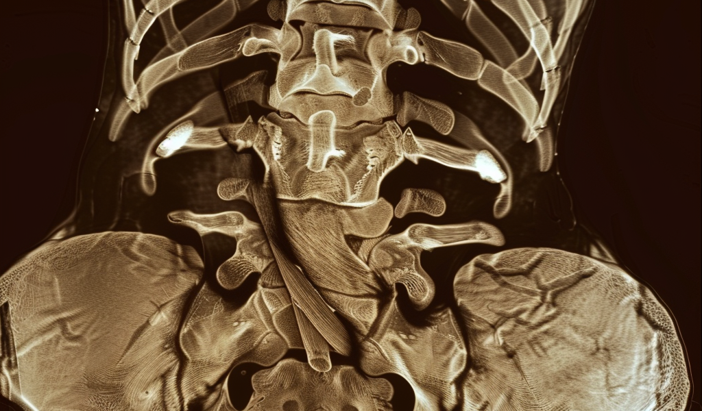
The study emphasizes the importance of targeting the appropriate patient population for CT utilization to enhance patient care. By analyzing CT scans for vertebral compression fractures through sagittal images, healthcare providers can detect and manage low bone mineral density more effectively.
As osteoporosis-related bone fractures are projected to increase significantly in China by 2050, costing billions of dollars, the need for accurate diagnostic tools is critical. While DEXA remains a common method for diagnosing osteoporosis, a large percentage of individuals with bone fractures do not undergo bone mineral density testing, and the efficacy of DEXA can be limited.
The researchers propose that evaluating bone mineral density using chest CT scans and measuring the Hounsfield unit (HU) expression in trabecular vertebrae could offer promising results. Chest CT scans, routinely used for lung cancer screening, can provide valuable insights into vertebral quality, particularly through sagittal images, which are essential for evaluating vertebral morphology.
In a study involving 1,268 patients who underwent both chest CT and DEXA scans, the researchers assessed the performance of CT in predicting low bone mineral density. The findings revealed that the diagnostic accuracy, as measured by the area under the receiver operating characteristic curve (AUC), was higher in women compared to men. Additionally, the AUC increased with age but decreased with height and weight.
For women, a CT attenuation value threshold of 140.25 HU showed a sensitivity of 69.3% in distinguishing low bone mineral density from normal, while in men, the sensitivity was 36.5%. Among individuals aged 70 or above, a threshold of 126.31 HU demonstrated a sensitivity of 76.1%, outperforming other age groups.
The study underscores the potential benefits of incorporating opportunistic chest CT scans in assessing bone health, particularly in populations where traditional methods may be limited. By leveraging CT imaging to evaluate bone mineral density, healthcare providers can enhance early detection and management of osteoporosis, ultimately improving patient outcomes.