Recent findings from the University of Rochester’s Del Monte Institute for Neuroscience have shed light on the structural differences in the brains of children with autism, paving the way for enhanced diagnostic methods and targeted treatment strategies. This groundbreaking study, published in the journal Autism Research, highlights significant variations in neuron density between children diagnosed with autism and their neurotypical counterparts, providing new insights into the neurological foundations of this complex disorder.
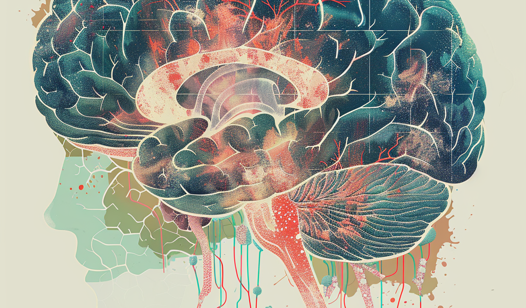
Led by Zachary Christensen, an MD/PhD candidate at the University of Rochester School of Medicine and Dentistry, the research team employed advanced neuroimaging techniques to delve into the intricacies of neuron structure. Their investigation revealed that children with autism exhibit lower neuron density in specific regions of the cerebral cortex that are crucial for memory, learning, and reasoning. In contrast, other areas such as the amygdala, which plays a vital role in emotional regulation, displayed an increased density of neurons.
Christensen remarked, “We’ve long studied the broader characteristics of brain regions—such as thickness and volume. But these newer neuroimaging techniques allow us to characterize cellular-level changes, revealing greater complexity in brain development, especially in children with autism.” This innovative approach enables researchers to move beyond traditional observations, offering a more nuanced understanding of how autism affects brain development.
The research team analyzed brain imaging data from over 11,000 children aged 9 to 11, sourced from the Adolescent Brain Cognitive Development (ABCD) study. Among these participants, 142 children had received an autism diagnosis. The study found that the neuron density differences were notably specific to children with autism when compared to those without neurodevelopmental disorders, as well as to children with common psychiatric conditions such as ADHD and anxiety. These findings bolster the validity of the research and its implications for future studies.
These discoveries not only illuminate the structural variances in the brains of children with autism but also suggest potential new avenues for diagnosis and treatment. Christensen explained the challenges faced in distinguishing autism from other co-occurring conditions, stating, “Autism is often accompanied by other diagnoses, such as anxiety, depression, or ADHD, making it difficult to distinguish what is specifically related to autism.” However, the new measurements of neuron structure may provide a reliable means to identify unique deviations associated with autism, which could lead to more personalized therapeutic interventions.
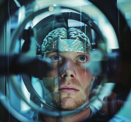
The advancements in neuroimaging technology have transformed the landscape of brain research, enabling scientists to explore neuronal architecture in living subjects rather than relying solely on postmortem examinations. This shift allows for the detection of subtle abnormalities that may have previously gone unnoticed, offering a clearer picture of how autism manifests in the brain.
As researchers continue to investigate these neuron density variations, the implications for autism diagnosis and treatment could be profound. The ability to pinpoint specific structural changes in the brain may facilitate earlier and more accurate diagnoses, ultimately leading to interventions that are tailored to the individual needs of each child.
In summary, the findings from the University of Rochester’s study represent a significant leap forward in our understanding of autism. By focusing on neuron density variations, researchers are uncovering vital information that could reshape the way autism is diagnosed and treated, providing hope for improved outcomes for children affected by this complex disorder.