New Research Highlights Role of Clec14a Protein in Bone Formation and Vascularization
Recent research has unveiled the critical function of the Clec14a protein in regulating bone formation and density, particularly in the context of capillary endothelial cells. This protein, identified as a type-H endothelial cell protein, plays a pivotal role in orchestrating osteoblast activity during the formation and patterning of trabecular bone.
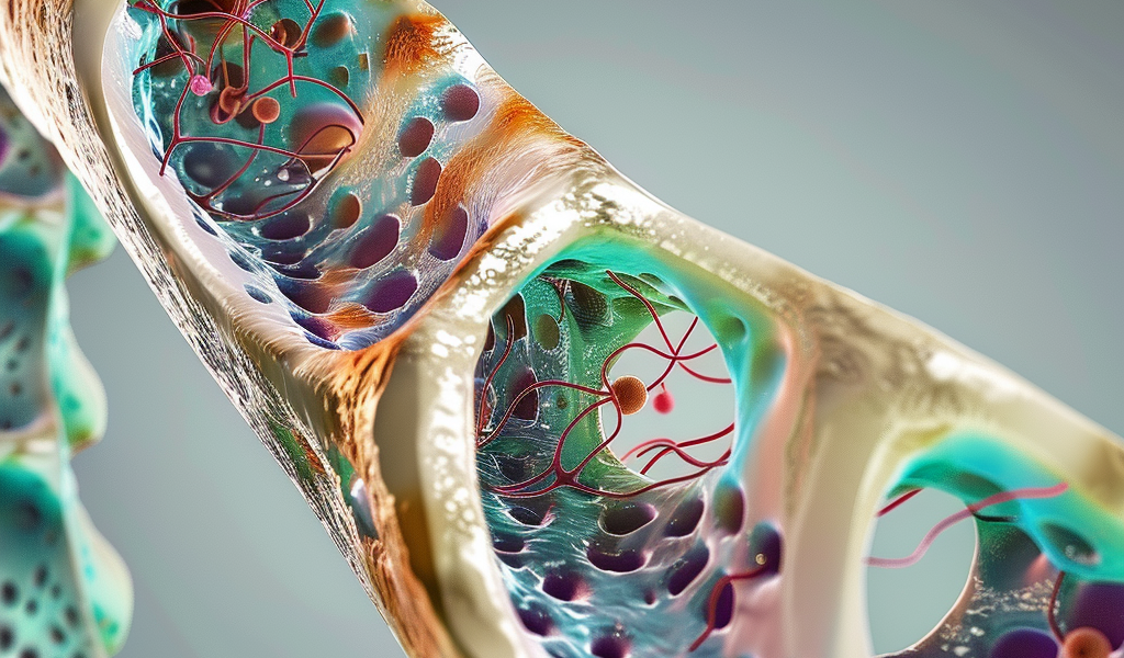
The study, published in Communications Biology, delves into the significance of Clec14a, formally known as C-type lectin domain containing 14A. It has garnered attention due to its involvement in bone angiogenesis, which is essential for postnatal skeletal growth, bone remodeling, and repair. Advances in technology have facilitated the discovery of new subtypes of bone capillaries, with type-H capillaries emerging as key players in modulating osteogenesis.
Another protein of interest, Multimerin 2 (MMRN2), is an extracellular matrix protein that binds to CD248 and CLEC14A. Both proteins are known to influence pathological and embryonic angiogenesis, as well as cell migration and adhesion. Previous studies indicated that CD248 negatively impacts osteoblast mineralization, and its deletion can enhance mineralization driven by osteoblasts. Additionally, there are indications that Clec14a expression increases during bone healing processes.
The researchers behind the current study aimed to further elucidate the role of Clec14a in bone formation. They began by examining the expression levels of Clec14a and its ligand, Mmrn2, in the long bones of mice. Their findings confirmed that both proteins were highly expressed in the bone metaphysis, a region critical for bone growth.
To visualize Clec14a in vivo, the researchers employed an anti-Clec14a antibody for immunolabeling. This technique revealed that Clec14a predominantly localizes to the type-H vessel endothelium within the bone metaphysis. The expression of Mmrn2 and Clec14a was found to be confined to endothelial cells (ECs), with a more pronounced expression in type-H vessel ECs.
Subsequently, the researchers investigated the effects of Clec14a deletion on bone vascularization. They compared two-week-old Clec14a-/- mice with wild-type (WT) Clec14a+/+ mice and noted a significant reduction in the percentage of type-H endothelium in the Clec14a-/- mice. Interestingly, both groups exhibited an increase in capillary networks from postnatal day four (P4) to four weeks of age.
Despite the differences in type-H endothelium, vessel density remained relatively similar between the two groups. Immunofluorescence imaging of the tibia at P4 showed a highly interconnected vascular bed, with comparable coverage of both type-L and type-H capillaries in both groups.
However, as the juvenile mice aged to four weeks, both groups displayed a noticeable decline in the type-H vascular bed compared to their earlier P4 state. This reduction was distinctly localized to the superior region of the metaphysis, highlighting the dynamic changes in vascularization that occur during bone development.
Overall, the study provides valuable insights into the role of Clec14a in bone health and development. Understanding the mechanisms by which Clec14a regulates osteoblast activity and bone vascularization could pave the way for new therapeutic strategies aimed at addressing bone-related disorders.